
Biology_A-level_Cie
-
1-1-the-microscope-in-cell-studies5 主题
-
1-2-cells-as-the-basic-units-of-living-organisms5 主题
-
2-1-testing-for-biological-molecules3 主题
-
2-2-carbohydrates-and-lipids8 主题
-
2-3-proteins6 主题
-
2-4-water2 主题
-
3-1-mode-of-action-of-enzymes5 主题
-
3-2-factors-that-affect-enzyme-action8 主题
-
4-1-fluid-mosaic-membranes4 主题
-
4-2-movement-into-and-out-of-cells12 主题
-
diffusion
-
osmosis
-
active-transport
-
endocytosis-and-exocytosis
-
investigating-transport-processes-in-plants
-
investigating-diffusion
-
surface-area-to-volume-ratios
-
investigating-surface-area
-
estimating-water-potential-in-plants
-
osmosis-in-plant-cells
-
osmosis-in-animals
-
comparing-osmosis-in-plants-and-animals
-
diffusion
-
5-1-replication-and-division-of-nuclei-and-cells6 主题
-
5-2-chromosome-behaviour-in-mitosis2 主题
-
6-1-structure-of-nucleic-acids-and-replication-of-dna4 主题
-
6-2-protein-synthesis5 主题
-
7-1-structure-of-transport-tissues4 主题
-
7-2-transport-mechanisms7 主题
-
8-1-the-circulatory-system7 主题
-
8-2-transport-of-oxygen-and-carbon-dioxide5 主题
-
8-3-the-heart4 主题
-
9-1-the-gas-exchange-system6 主题
-
10-1-infectious-diseases3 主题
-
10-2-antibiotics3 主题
-
11-1-the-immune-system4 主题
-
11-2-antibodies-and-vaccination6 主题
-
12-1-energy5 主题
-
12-2-respiration11 主题
-
aerobic-respiration-the-krebs-cycle
-
aerobic-respiration-role-of-nad-and-fad
-
aerobic-respiration-oxidative-phosphorylation
-
anaerobic-respiration
-
energy-yield-aerobic-and-anaerobic-respiration
-
anaerobic-adaptation-of-rice
-
aerobic-respiration-effect-of-temperature-and-substrate-concentration
-
structure-and-function-of-mitochondria
-
the-four-stages-in-aerobic-respiration
-
aerobic-respiration-glycolysis
-
aerobic-respiration-the-link-reaction
-
aerobic-respiration-the-krebs-cycle
-
13-1-photosynthesis-as-an-energy-transfer-process8 主题
-
13-2-investigation-of-limiting-factors2 主题
-
14-1-homeostasis-in-mammals8 主题
-
14-2-homeostasis-in-plants3 主题
-
15-1-control-and-coordination-in-mammals12 主题
-
the-endocrine-system
-
the-nervous-system
-
neurones
-
sensory-receptor-cells
-
sequence-of-events-resulting-in-an-action-potential
-
transmission-of-nerve-impulses
-
speed-of-conduction-of-impulses
-
the-refractory-period
-
cholinergic-synapses
-
stimulating-contraction-in-striated-muscle
-
ultrastructure-of-striated-muscle
-
sliding-filament-model-of-muscular-contraction
-
the-endocrine-system
-
15-2-control-and-coordination-in-plants3 主题
-
16-1-passage-of-information-from-parents-to-offspring5 主题
-
16-2-the-roles-of-genes-in-determining-the-phenotype7 主题
-
16-3-gene-control3 主题
-
17-1-variation4 主题
-
17-2-natural-and-artificial-selection7 主题
-
17-3-evolution2 主题
-
18-1-classification5 主题
-
18-2-biodiversity7 主题
-
18-3-conservation6 主题
-
19-1-principles-of-genetic-technology11 主题
-
19-2-genetic-technology-applied-to-medicine4 主题
-
19-3-genetically-modified-organisms-in-agriculture2 主题
-
1-1-the-microscope-in-cell-studies
-
1-2-cells-as-the-basic-units-of-living-organisms
-
2-1-testing-for-biological-molecules
-
2-2-carbohydrates-and-lipids
-
2-3-proteins
-
2-4-water
-
3-1-mode-of-action-of-enzymes
-
3-2-factors-that-affect-enzyme-action
-
4-1-fluid-mosaic-membranes
-
4-2-movement-into-and-out-of-cells
-
5-1-replication-and-division-of-nuclei-and-cells
-
5-2-chromosome-behaviour-in-mitosis
-
6-1-structure-of-nucleic-acids-and-replication-of-dna
-
6-2-protein-synthesis
-
7-1-structure-of-transport-tissues
-
7-2-transport-mechanisms
-
8-1-the-circulatory-system
-
8-2-transport-of-oxygen-and-carbon-dioxide
-
8-3-the-heart
-
9-1-the-gas-exchange-system
-
10-1-infectious-diseases
-
10-2-antibiotics
-
11-1-the-immune-system
-
11-2-antibodies-and-vaccination
eyepiece-graticules-and-stage-micrometers
Eyepiece graticules & stage micrometers
-
An eyepiece graticule and stage micrometer are used to measure the size of an object when viewed under a microscope
-
The eyepiece graticule is an engraved ruler that is visible when looking through the eyepiece of a microscope
-
Eyepiece graticules are often divided into 100 smaller divisions known as graticule divisions, or eyepiece units
-
-
The values of the divisions in an eyepiece graticule vary depending on the magnification used, so the graticule needs to be calibrated every time an object is viewed
-
The calibration is done using a stage micrometer; this is a slide that contains a tiny ruler with an accurate known scale
-
Stage micrometer rulers can vary, but often have larger divisions of 0.1 mm (100 μm) and smaller divisions of 0.01 mm (10 μm)
-
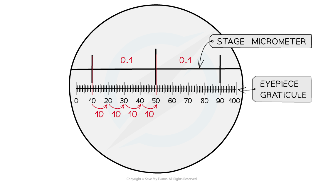
Calibrating the eyepiece graticule
-
In the diagram, two stage micrometer divisions of 0.1 mm, or 100 μm, are visible
-
Each 100 µm division is equal to 40 eyepiece graticule divisions
-
40 graticule divisions = 100 µm
-
1 graticule division = number of µm ÷ number of graticule divisions
-
1 graticule division = 100 ÷ 40 = 2.5 µm; this is the magnification factor
Calculating the size of a specimen
-
The calibrated eyepiece graticule can be used to measure the length of an object
-
The number of graticule divisions covered by an object need to be multiplied by the magnification factor:
Graticule divisions covered by object x magnification factor = length of object (µm)
Worked Example
A student viewed some onion cells under a microscope.
The image below shows the cells with an eyepiece graticule (left) and the eyepiece graticule alongside a stage micrometer (right).
Note that each large division on the stage micrometer here is 100 μm, and each small division is 10 μm.
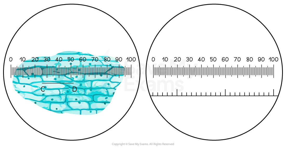
Use the stage micrometer to calibrate the eyepiece graticule and calculate the actual length of the cell labelled C-D in the image.
Answer:
Step 1: Calculate the size of each eyepiece division
There are 40 graticule divisions per large micrometer division, or per 100 μm
1 graticule division = no. of μm ÷ no. of graticule divisions
= 100 ÷ 40
= 2.5 μm
This value can now be used as a magnification factor
Step 2: Calculate the length of the cell
Specimen size = no. of graticule divisions x magnification factor
The cell closest to the ruler covers 27 graticule divisions
= 27 x 2.5
= 67.5 μm
Step 3: Consider whether this answer makes sense in context
Plant cells usually measure between 10-100 μm, so a result of 67.5 μm sounds sensible in this context
Examiner Tips and Tricks
The calculations involving stage micrometers and eyepiece graticules are often seen in exam questions, so make sure that you are comfortable with how to calibrate the graticule and calculate the length of an object on the slide.
Note that both of the examples given above are carried out at the same magnification, so the magnification factor calculated during calibration is 2.5 in both cases. This may not always be the same in an exam.